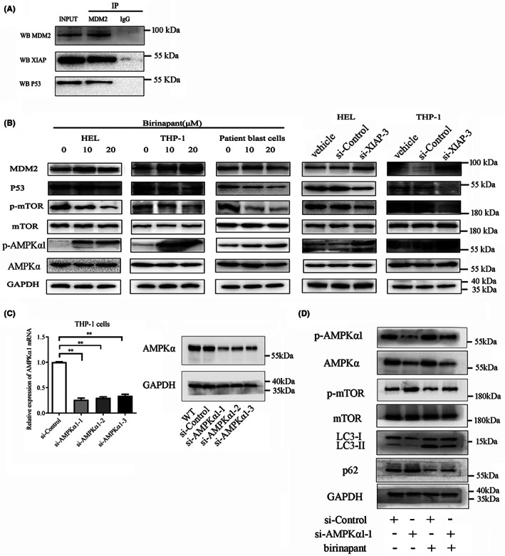
FIGURE 5.

XIAP inhibition promotes autophagy through MDM2‐p53‐AMPK pathway. (A) Co‐IP showed that XIAP interacted with MDM2 and p53 in HEL cells. Ten percent whole cell lysate as INPUT samples was used. (B) XIAP inhibition using birinapant or si‐XIAP‐3 downregulated the level of p53 and the phosphorylation of mTOR, upregulated the phosphorylation of AMPK, but almost unaffected the level of MDM2 in AML cells. (C) siRNAs were designed to knockdown AMPKα1 and its effects were verified by relative expression of AMPKα1 mRNA and protein using qRT‐PCR and western blot in THP‐1 cells. (D) THP‐1 cells were transfected with si‐AMPKα1‐1 or si‐Control for 24 h, subsequently treated either alone or in combination with 20 μM birinapant for 48 h, and then, the cells were collected and determined for the expression of p‐AMPKα1, AMPKα1, p‐mTOR, mTOR, LC3‐I/II, p62 and GAPDH using western blot, respectively. Images shown were representatives of at least three independent experiments.
