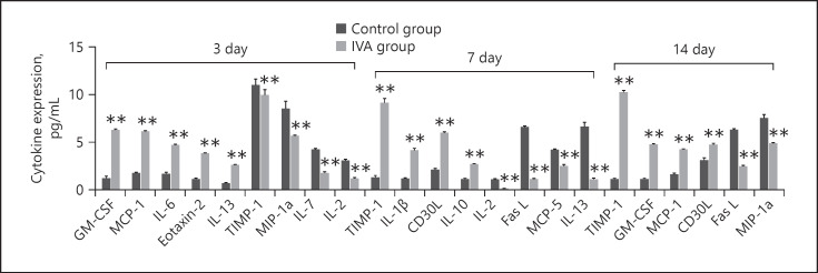
Fig. 2.
Differentially expressed cytokines between CNV groups and aflibercept treatment groups on days 3, 7, and 14 measured using a multiplex quantitative cytokines array (p < 0.001, fold change >2.0). GM-CSF and MCP-1 were the most significantly upregulated cytokines on day 3 after laser induction (p < 0.001, fold change >5.0). MIP-1a was the most significantly downregulated cytokine on day 3 after laser induction (p < 0.001, fold change >5.0). TIMP-1 was the most significantly upregulated cytokine on day 7 after laser induction (p < 0.001, fold change >5.0). IL-13 and Fas-L were the most significantly downregulated cytokines on day 7 after laser induction (p < 0.001, fold change >5.0). TIMP-1 was the most significantly upregulated cytokine on day 14 after laser induction (p < 0.001, fold change >5.0). Fas-L was the most significantly downregulated cytokine on day 14 after laser induction (p < 0.001, fold change >5.0). Data are presented as mean + SEM, n = 3 for each time point. **p < 0.001 versus control. CNV, choroidal neovascularization; GM-CSF, granulocyte-macrophage colony-stimulating factor; MIP, macrophage inflammatory protein; MCP, monocyte-chemoattractant protein; Fas-L, Fas ligand; IL, interleukin; TIMP, tissue inhibitor of metalloproteinase.

