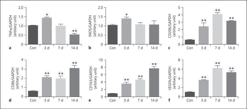
Fig. 3.
RT-PCR analysis of gene expression of TNF-α, iNOS, CD206, CD86, CD163, and VEGFA in the normal RPE-choroid-sclera complex and that of laser-induced CNV mice model at different time points. The level of TNF-α increased on day 3 after laser photocoagulation, while significantly decreased on day 14 after laser photocoagulation (a). The level of iNOS increased on day 3 after laser photocoagulation (b). The level of CD206 increased on days 3, 7, and 14 after laser photocoagulation and peaked on day 7 (c). The level of CD86 (d) and CD163 (e) increased on days 3, 7, and 14 after laser photocoagulation and peaked on day 14. The level of VEGFA increased on days 3, 7, and 14 after laser photocoagulation and peaked on day 7 (f). Data are presented as mean ± SEM, n = 6 for each time point. *p < 0.05, **p < 0.001.

