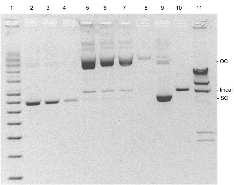
Figure 1.

Agarose gel electrophoresis of supercoiled circular (SC), open circular (OC) and linear forms of pSVβ. Lane 1, supercoiled DNA ladder; lanes 2–4 and 9, 105, 61.5, 17.5 and 350 ng, respectively, of a typical plasmid preparation (P-DNA); lanes 5–8, 350, 105, 61.5 and 17.5 ng, respectively, of the same preparation but incubated at 60°C for 48 h (O-DNA); lane 10, P-DNA linearized with HindIII; lane 11, phage λ × HindIII molecular weight marker.
