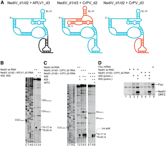
FIGURE 8.
80S complex formation on chimeric NediV/APLV1 and NediV/CrPV IRESs. (A) Schematic models of hybrid IRESs. Elements derived from NediV, APLV1, and CrPV IRESs are colored light blue, black, and red, respectively. (B) Binding of ribosomes to NediV wt and NediV/APLV1 chimeric IRESs, assayed by toeprinting. Separation of lanes by white lines indicates that they were juxtaposed from the same gel. (C) Binding of 40S or 40S and 60S subunits to NediV wt and NediV/CrPV chimeric IRESs, followed by elongation on inclusion of eEF1H/2H, ∑aa-tRNA, and cycloheximide (CHX), assayed by toeprinting. (B,C) Positions of the P site codon and of bound ribosomal complexes are indicated. Lanes C, T, A, G depict NediV sequence.

