
Figure 5.
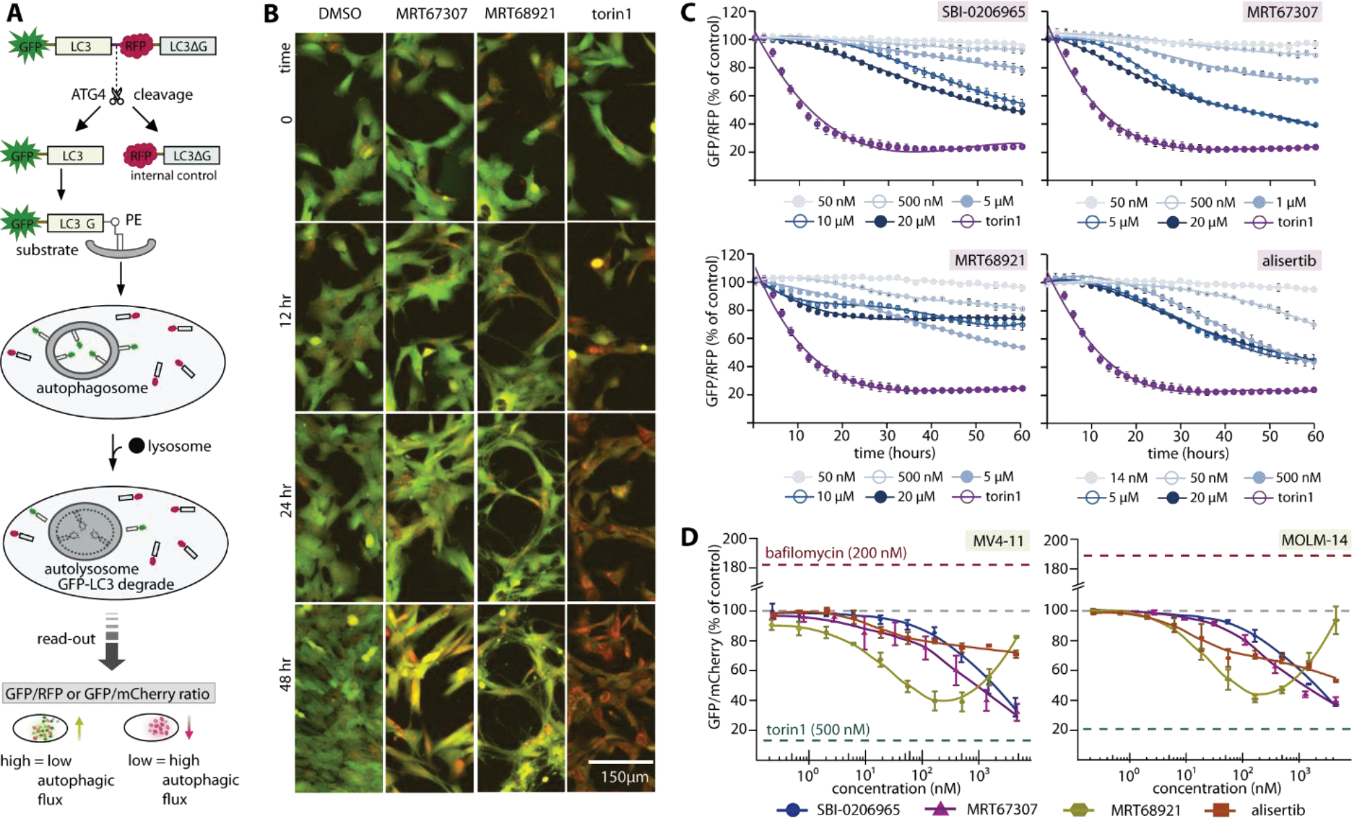
Complex cellular effects of ULK1/2 inhibitors on autophagy modulation due to off-target effects. A) Schematic diagram illustrates LC3-based GFP/RFP fluorescence autophagy flux assays used in RPE1 experiment. A similar assay scheme was used in leukemia cell lines, albeit with an omission of C-terminal LC3ΔG from the GFP-LC3B-mCherry probe. The read-out GFP/RFP or GFP/mCherry ratio from high to low confers low to high autophagy flux levels, respectively. B) Examples of RPE1 cells at different time point in the autophagic flux assays. Fluorescent images (overlay green and red) of RPE1 cells stably expressing the autophagy flux reporter GFP-LC3-RFP-LC3ΔC. Cells were treated for the indicated time with 1% DMSO, ULK1 inhibitor MRT67307 and MRT68921 at 5 μM, and 250 nM torin1, respectively. A drop in GFP (green channel) indicates increased autophagy flux. C) Time-dependent progression of autophagic flux in RPE1 cells of three ULK1 inhibitors and selective Aurora A inhibitor alisertib at various concentrations (torin1 used as control at 250 nM). D) Autophagic flux measured at 24-hour time point reveals dose-response of autophagic flux of the same set of inhibitors in leukemia cell line MV4-11 and MOLM-14. For both (C) and (D), mean of repeated experiment with standard error of the mean (SEM) are shown.