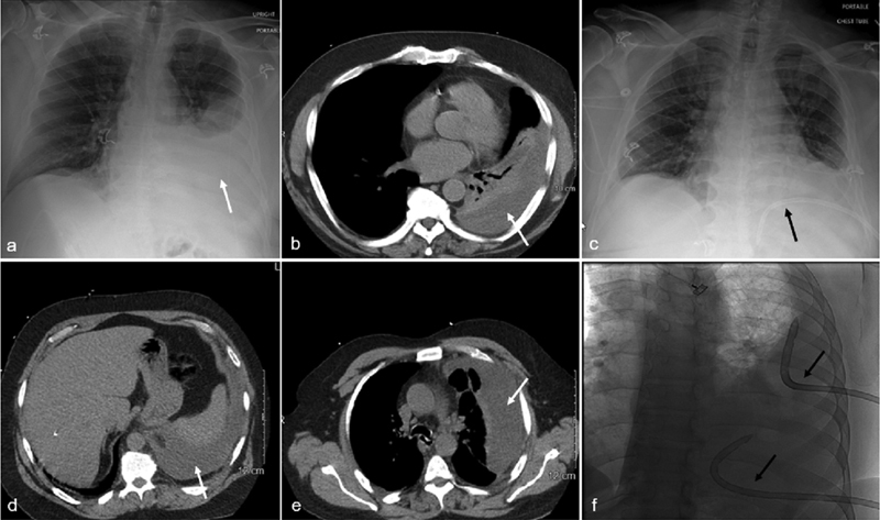
Fig. 1.

Anatomic positioning of chest tubes. A 65-year-old male with a loculated left parapneumonic pleural effusion. Chest radiograph ( a ) and CT of the chest ( b ) show the posterior pleural fluid collection (white arrow). A 16-Fr Gordon angled chest tube (white arrow) is placed into the dependent posterior inferior left pleural space (black arrow) ( c ). A 55-year-old male with Staphylococcus and Bacteroides empyema. CT of the chest ( d, e ) shows a large, loculated left pleural collection (white arrow). Due to the size and extent of loculations of the empyema, two chest tubes are placed to efficiently drain the infected fluid along with intracatheter lytic therapy. A fluoroscopic image ( f ) depicts an 18-Fr Gordon angled chest tube in the superior lateral left pleural space and another tube in the posterior inferior left pleural collection (black arrows).
