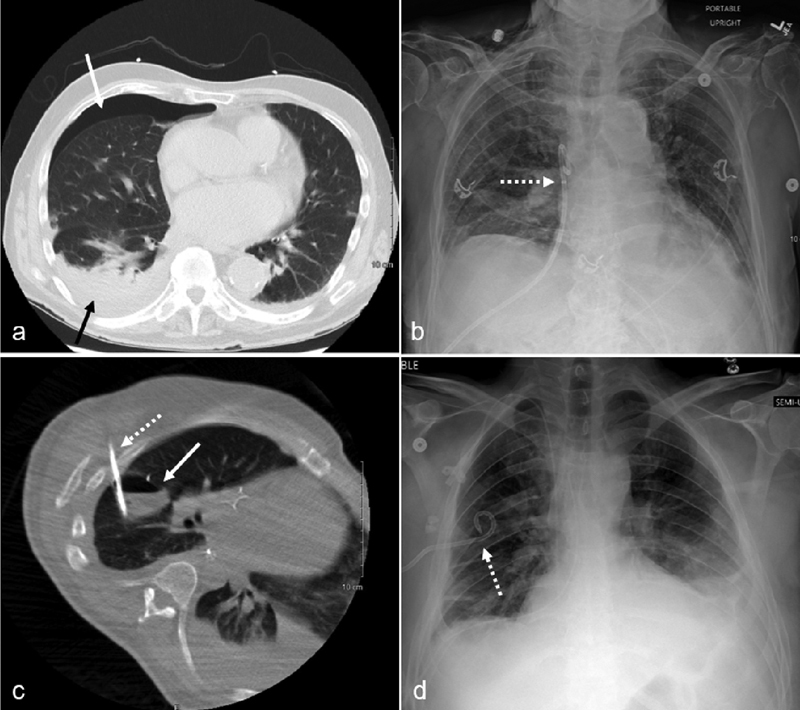
Fig. 2.

An 85-year-old male presented with a delayed presentation of a right pneumothorax following parathyroid surgery. Chest CT ( a ) demonstrates a small right pneumothorax (solid white arrow), small pleural effusion (solid black arrow), and basilar atelectasis. A 12-Fr, nonlocking pigtail catheter (dotted arrow) was placed into the anterior right pleural space using CT guidance as seen on the subsequent chest radiography ( b ). A 38-year-old male with an infected hydropneumothorax following an Ivor-Lewis esophagectomy. Chest CT during drainage procedure (c) shows a loculated gas and fluid collection in the right major fissure (solid white arrow). Using CT guidance, a 12-Fr locking pigtail chest tube (dotted white arrows) was placed into the intrafissural collection as demonstrated by CT (c) and chest radiograph ( d ).
