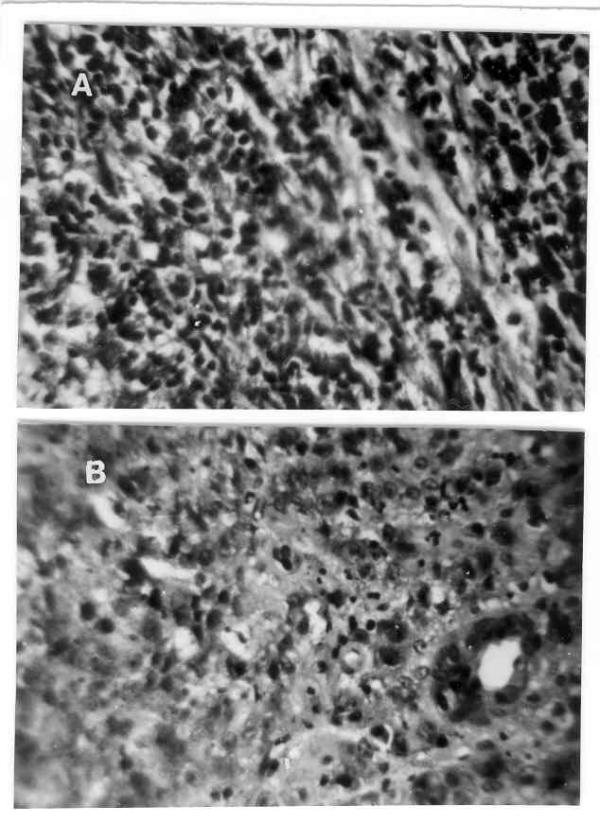
Figure 2.

(A) Resected specimen of distal stomach showing diffuse infiltration by mononuclear cells without formation of lymphoid follicles with obvious cellular atypia and abnormal mitotic figures (H&E × 275). (B) The high power view of recurrent gastric tumor showing pleomorphic cells, abnormal mitotic figures, mucin secretion and formation of gland at places diagnostic of adenocarcinoma (H&E × 275).
