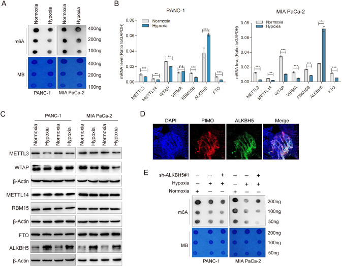
Fig. 1. Hypoxia decreased m6A modification depended on ALKBH5 in pancreatic cancer.
A PANC-1 and MIA PaCa-2 cells were exposed to either 20% or 1% O2 for 48 h. mRNA was extracted and m6A levels were determined by dot blot. RNAs were serially diluted and loaded equally with the amount of 400 ng, 200 ng and 100 ng. The intensity of dot immunoblotting (up) represented the level of m6A modification. MB, methylene blue staining (down) as loading control. B qRT-PCR assays were performed to analyze the mRNA levels of METTL3, WTAP, METTL14, RBM15, FTO and ALKBH5 in indicated PANC-1 and MIA PaCa-2 cells grown for 48 h under normoxia or hypoxia. C PANC-1 and MIA PaCa-2 cells were exposed to either 20% or 1% O2 for 48 h. Immunoblots showing the protein levels of METTL3, WTAP, METTL14, RBM15, FTO and ALKBH5. n = 3 independent experiments. D Immunofluorescence staining of ALKBH5 and pimonidazole (PIMO) in PANC02-derived mouse tumors. Hypoxic tumor areas were marked by PIMO staining. E Dot blot assays were used to measure m6A levels in sh-NC and shALKBH5 PC cells exposed to either 20% or 1% O2 for 48 h. Error bars indicate means ± SEM, n = 3, *P < 0.05, **P < 0.01, ***P < 0.001 and ns not significant.

