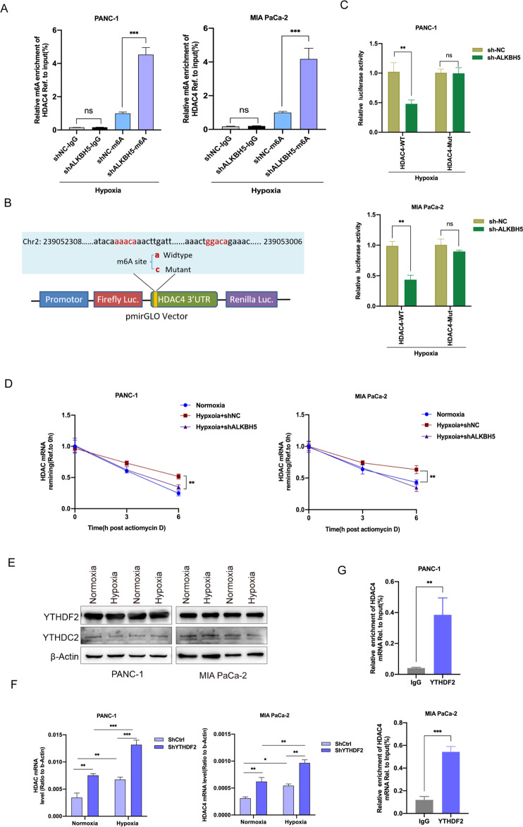
Fig. 4. HDAC4 expression was regulated by m6A modification.
A Reduction in m6A modification in specific regions of HDAC4 transcripts upon ALKBH5 knockdown in PANC-1 and MIA PaCa-2 cells under hypoxia for 48 h by gene-specific m6A-RIP-qPCR assays. The value obtained for the control group was set to 1. B Graphical explanation for construction of luciferase reporters. The wild-type or mutant of m6A motif sequence of HDAC4 3’UTR was inserted into a pcDNA3.1 vector. C Dual luciferase reporter assays showing the effect of ALKBH5 on HDAC4 mRNA reporters with either wild-type or mutated m6A sites under hypoxia. D The stability of HDAC4 in ALKBH5 knockdown and its corresponding control PC cells. Cells were treated with actinomycin D (2 µg/mL) at the indicated time points (0, 3 and 6 h) and were detected by qRT-PCR. Error bars are mean ± SEM. (n = 3). E Western blot showing YTHDF2 and YTHDC1 protein levels in normoxic or hypoxic PC cells. F The expression level of HDAC4 in YTHDF2 knockdown and its corresponding control PC cells with or without hypoxia were detected by qRT-PCR. Error bars are mean ± SEM. (n = 3). G The combination of HDAC4 and YTHDF2 was detected by RIP-qPCR analysis using anti-IgG and anti-YTHDF2 antibodies. Error bars are mean ± SEM. (n = 3). *P < 0.05, **P < 0.01, ***P < 0.001 and ns not significant.

