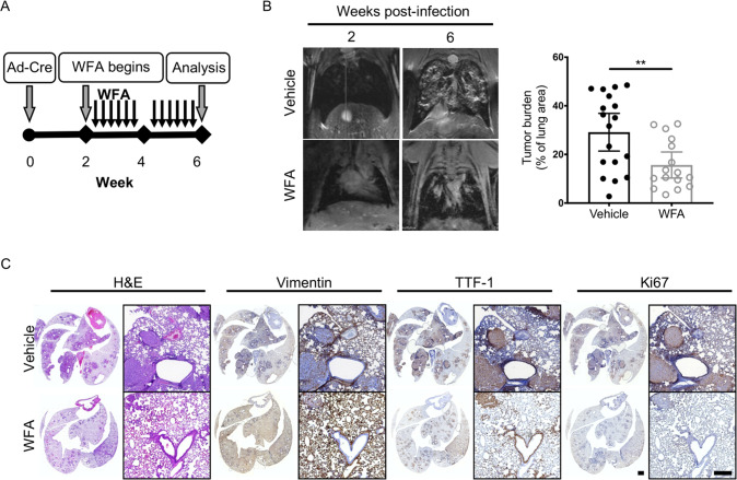
Fig. 4. WFA treatment attenuates lung cancer progression.
A Schematic of experimental design. KPV+/+ mice were treated with withaferin A (WFA; 4 mg/kg; QOD, p.o.) or vehicle control (DMSO) at 2 weeks post-infection with 107 PFUs of adenoviral Cre. B Representative MRI scans show WFA-treated KPV+/+ lung tumors at 6 weeks post-infection with 107 PFUs of adenoviral Cre (left). Dot plot illustrates the tumor volume between WFA-treated or vehicle-treated control KPV+/+ mice (right). Each point represents, for one mouse, the percentage of lung area on MRI occupied by tumor, as measured using Jim software. Data are presented as the mean ± standard deviation (**p < 0.01 by unpaired, two-tailed t-test). C Lungs isolated from vehicle- or WFA-treated KPV+/+ mice 6 weeks after adenoviral Cre infection were fixed, sectioned, and subjected to H&E staining and vimentin, TTF-1, and Ki67 immunohistochemical staining. Positively immunostained cells appear brown, and nuclei are dyed blue. Scale bars: 2 mm (whole lungs, left), 200 µM (right).

