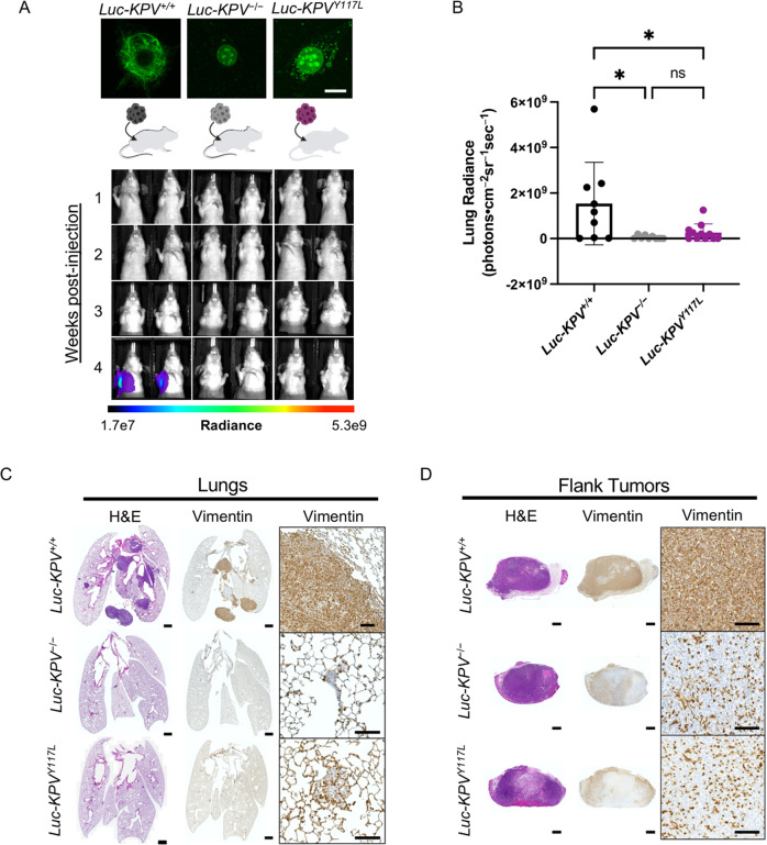
Fig. 6. Vimentin is required for accelerated lung cancer metastasis.
A (Top) Luc-KPV−/− cells were transfected with vimentin-Y117L to create Luc-KPVY117L cells. Cells were stained with DAPI and an anti-vimentin antibody (green). Scale bar: 10 µm. (Bottom) A total of 1 × 10⁶ KPV+/+, KPV−/−, or KPVY117L cells labeled with luciferase (Luc-KPV+/+, Luc-KPV−/−, and Luc-KPVY117L, respectively) were injected subcutaneously into the right flank of nude mice. At 3 weeks post-injection, primary tumors were removed and lung metastases were tracked for an additional 1 week. Shown are representative IVIS images of mice (n = 9–11 per group). The coronal views shown were acquired after masking the flank tumor to minimize bleed-through of the signal. Intensity overlay shows the accumulation of luciferase-labeled cells. B Luciferin signal was quantified from the lungs. An ordinary one-way ANOVA with multiple comparisons was used to compare groups at week 4 (*p < 0.05). C Lungs at week 4 and D excised flank tumors from week 3 were fixed, sectioned, and subjected to H&E staining and vimentin immunohistochemical staining. Positive vimentin staining is brown, and nuclei are blue. Scale bars: 1 mm (whole tumor/lung, left), 100 μm (inset, right).

