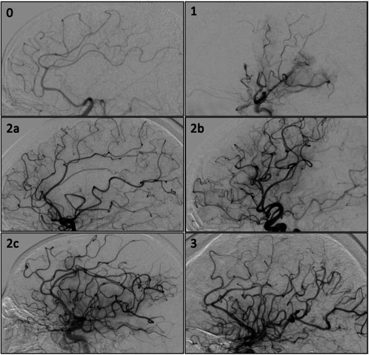
Fig. 1.
Recanalization assessment on digital subtraction angiography using the expanded Thrombolysis in Cerebral Infarction (eTICI) score. The images show lateral intracranial angiograms after intra-arterial contrast injection into the internal carotid artery. eTICI 0—no vessels in the middle cerebral artery territory are opacified. eTICI 1—only very limited opacification of the proximal middle cerebral artery, with no filling of any distal branches is seen. eTICI 2a—normal vascular opacification in < 50% of the middle cerebral artery territory. eTICI 2b—normal vascular opacification in 50–90% of the middle cerebral artery territory. eTICI 2c—normal vascular opacification in 90–99% of the middle cerebral artery territory. eTICI 3—normal vascular opacification in the complete middle cerebral artery territory (100%)

