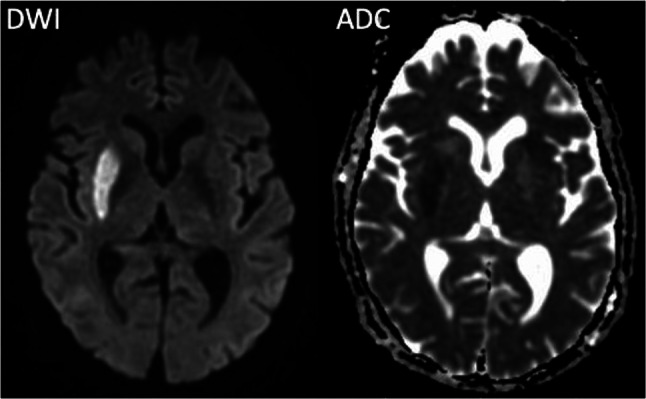
Fig. 4.

Diffusion-weighted (DWI) MRI and corresponding apparent diffusion coefficient (ADC) maps. Areas with cytotoxic edema, i.e., areas with insufficient tissue perfusion that are infarcted or invariably going to infarct, can be seen as hyperintense signal on DWI with a corresponding decrease (hypointense signal) on the ADC map. The image pair shows a right-sided acute infarct in the lentiform nucleus
