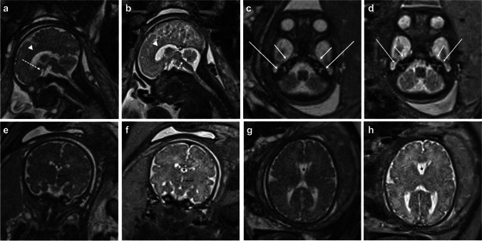
Fig. 1.
34-week gestational age fetus. Sagittal, axial, and coronal representative images of the fetal brain with half-Fourier single-shot turbo spin echo (HASTE) and balanced steady-state free precession (bSSFP). Sagittal bSSFP (a) and HASTE (b) images show normal midline structures including the corpus callosum (arrowhead), optic chiasm/nerve (long dashed arrow), and 4th ventricle (short dashed arrow). Axial bSSFP (c) and HASTE (d) images at the level of the inner ears partially resolves the cochlea (short solid arrow) and semi-circular canals (long solid arrow). Coronal bSSFP (e) and HASTE (f) images through the frontal horns demonstrate a normal cavum septi pellucidi (star), third ventricle (black arrow), and normal sulcal/gyral pattern. Axial bSSFP (g) and HASTE (h) images through the lateral ventricles show a normal cavum septi pellucidi (star), lateral ventricles, and normal sulcal/gyral pattern

