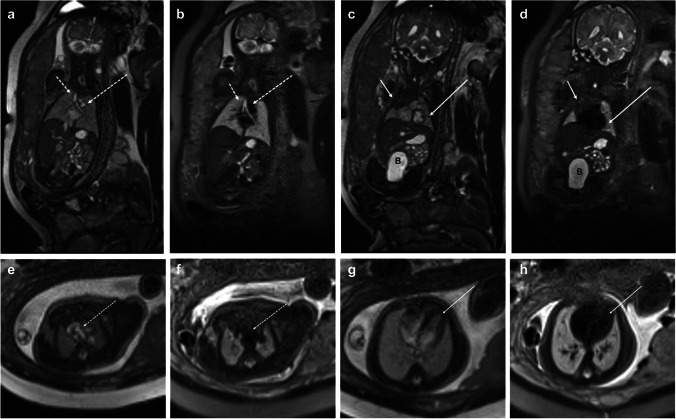
Fig. 2.
32-week gestational age fetus. Coronal balanced steady-state free precession (bSSFP) (a) and half-Fourier single-shot turbo spin echo (HASTE) (b) images show a normal fluid-filled tracheobronchial tree (short dashed arrow), mediastinal vascular structures (long dashed arrow), liver, stomach, and bowel. Coronal bSSFP (c) and HASTE (d) images more anterior demonstrate a normal thymus (short solid arrow) and left ventricle (long solid arrow), and bladder (B) as well. Axial bSSFP (e) and HASTE (f) images at the level of the great vessels and axial bSSFP (g) and HASTE (h) show bright and black blood images of flowing blood into the heart (long solid arrow) and great vessels (long dashed arrow) and contrast with the fluid-filled lungs

