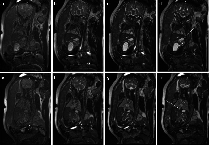
Fig. 5.
34-week gestational age fetus. Coronal whole body balanced steady-state free precession images at increasing flip angles, 30° (a, e), 60° (b, f), 90° (c, g), and 120° (d, h). As flip angles increased, there was improved signal to noise and contrast resolution, in particular flowing blood in the inferior vena cava (long dashed arrow) versus fluid filled lungs (short dashed arrow) versus static fluid in the stomach (long solid arrow) and bladder (short solid arrow)

