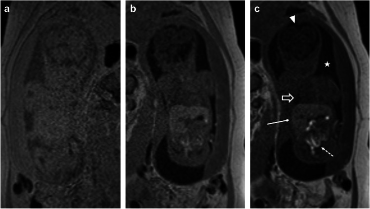
Fig. 6.
25-week gestational age fetus. Coronal spoiled gradient-recalled echo images at increasing flip angles, 15° (a), 30° (b), and 60° (c), show improved contrast of short T1 structures such as liver (long solid arrow) and meconium filled bowel (short dashed arrow), as well as improved but subtle gray-white matter differentiation. Additionally, there is increased distinction of tissues with longer T1, such as amniotic fluid (star), fluid-filled lungs (open arrow), and cerebral spinal fluid (arrowhead)

