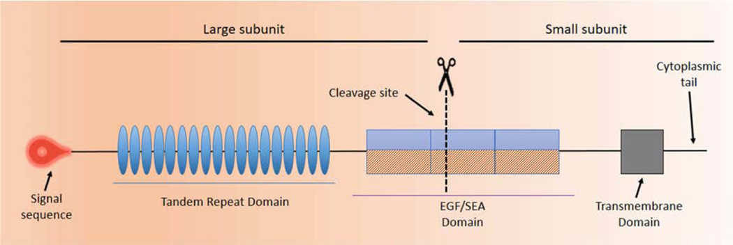
Figure 4. Prototype of a Membrane Associated Mucin (MAM).
The graphic depicts a prototypical MAM, the structure of which is similar to a classic, single-pass transmembrane immune receptor. A signal peptide motif is found at the N-terminal of the precursor polypeptide chain to enable its membrane insertion; it may be retained in the mature protein (1). The mature protein is composed of two subunits that self-associate, arising from intracellular cleavage. The large subunit is entirely extracellular and contains the VNTR. The small subunit consists of a short extracellular region, a single-pass transmembrane domain, and a cytoplasmic tail (CT). The large subunit of the MAM, together with the extracellular portion of the small subunit, comprise the extracellular domain (ED). The ED also contains conserved sequence motifs as modular elements such as the Sperm protein, Enterokinase and Agrin module (SEA) and EGF-like modules.

