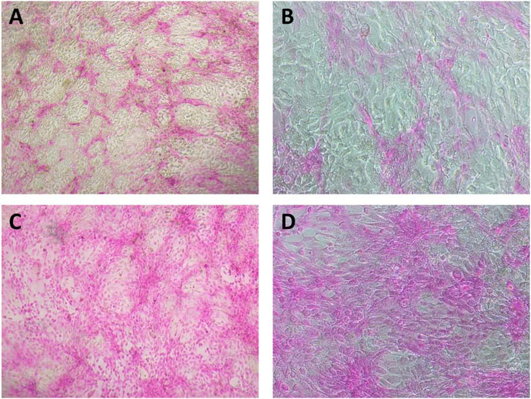
Fig 8. Application of Oxidative Stress to HCLE Epithelial-Equivalents with Mucosal Differentiation.

Islands of cells with mucosal differentiation at the surface of HCLE epithelial-equivalents exclude rose bengal (A and B). When oxidative stress is applied (10 mM tBHP in DMEM/F12 medium for 2 hours, as previously described (Webster et al., 2018)), many of these cells lose their transcellular barrier function and rose bengal penetrates (C and D). Rose Bengal staining was performed by incubating the cells 5 minutes in 0.1% Rose Bengal as previously described (Argueso et al., 2006). Magnification: A and C are 4x; B and D are 10x.
This example has not been previously published, but is similar to findings published in (Webster et al., 2018)
