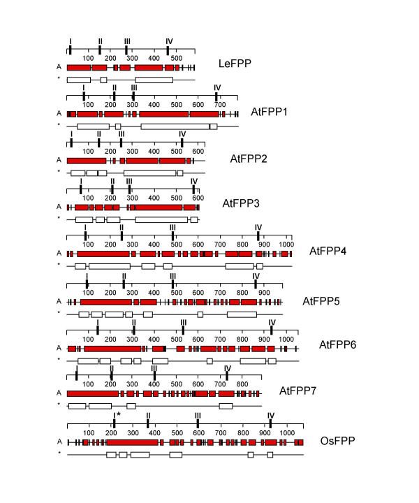
Figure 3.
Comparison of predicted secondary structure of FPP proteins. All proteins are drawn to scale. The numbers on the scales represent amino acids. Alpha helices are indicated as gray bars (A); coiled-coil domains are indicated as white bars (*). The positions of the four conserved sequence motifs are indicated as I, II, III, and IV on the scales. I* indicates the variant of motif I found in OsFPP.

