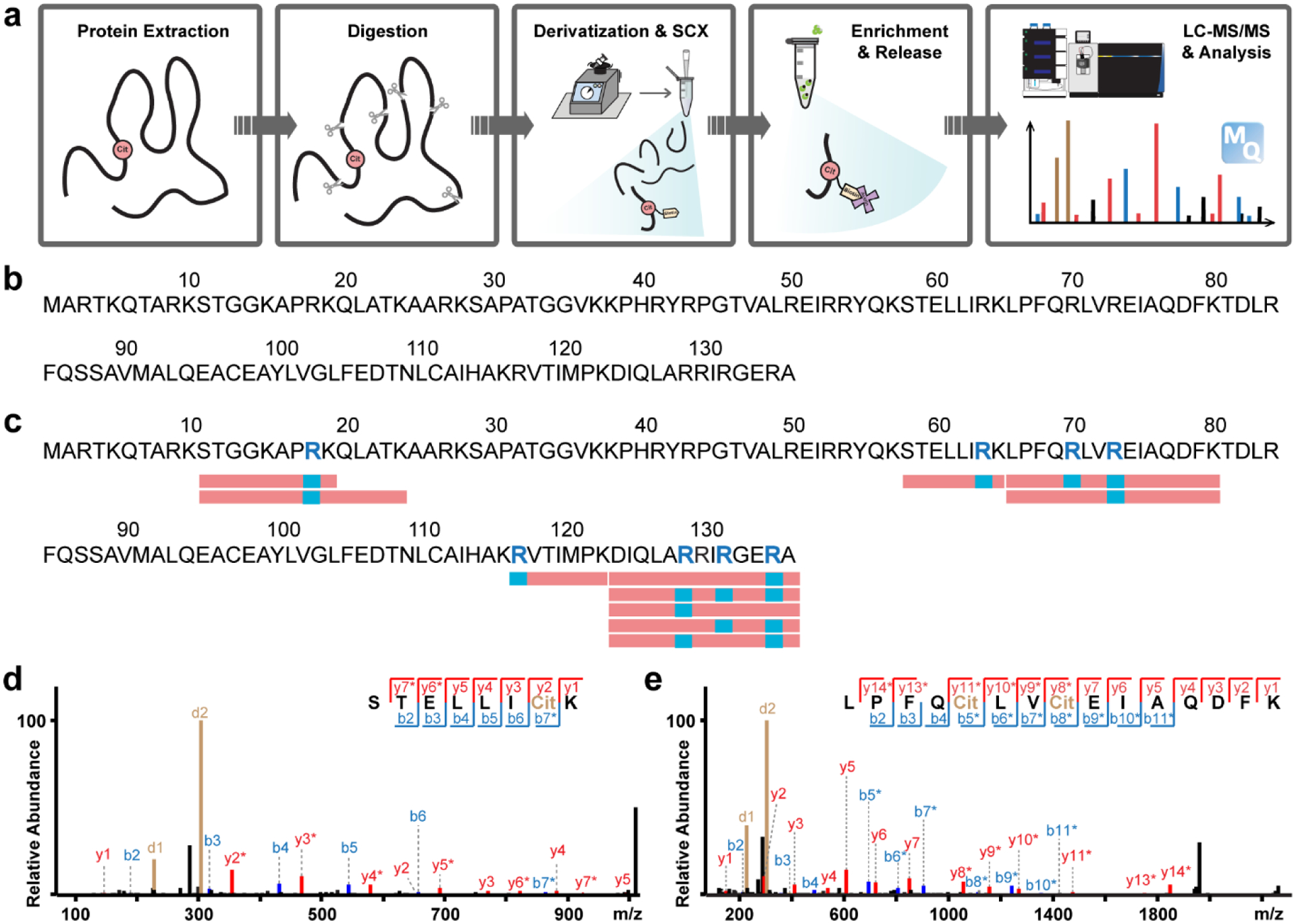
Figure 2. Improved in vitro protein citrullination analysis with biotin thiol tag.

a, Experimental workflow of protein citrullination analysis with biotin thiol tag. b,c, Citrullination analysis of histone H3 protein before (b) and after (c) in vitro PAD treatment. Identified citrullination sites are highlighted as blue letters within the sequence. Red rectangles below the sequence indicate confidently identified citrullinated peptides while citrullination sites are shown in blue. d, Example tandem MS spectrum of an identified citrullinated peptide from PAD treated histone H3 (R64Cit). e, Example tandem MS spectrum of two citrullination sites (R70Cit and R73Cit) identified on the same peptide from PAD treated histone H3.
