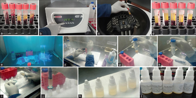
Figure 2.
Illustrative collage of images describing the process of preparing 20% autologous serum eye drops. (a) Blood collected in sterile plain vacutainers and kept at room temperature (30 minutes) for clotting; (b and c) Centrifugation of the tubes for serum separation; (d) Well-separated serum seen in the upper part of the tubes with RBC compacted at the bottom; (e) Laminar flow hood with UV light exposure (30 minutes) to sterilize all items required for serum separation, dilution, and packaging; (f) Serum being pipetted out in a sterile tube; (g and h) Sterile normal saline being added to the serum for dilution; (i and j) Millipore syringe filter of 0.22 μm being fixed to a syringe containing the diluted serum; (k) 5 mL of sterile diluted serum delivered into sterile eye dropper vials (note: few drops of the diluted serum is placed in culture media at this stage); (l) Appropriate label fixed on the eye dropper vials with relevant details

