Figure 2.

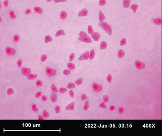
Photomicrograph of impression cytology specimen, stained with periodic acid-Schiff, and hematoxylin-eosin at X 400 with squamous metaplasia. Showing both normal cells (NC) and increased nuclear–cytoplasmic ratio (SM)

Photomicrograph of impression cytology specimen, stained with periodic acid-Schiff, and hematoxylin-eosin at X 400 with squamous metaplasia. Showing both normal cells (NC) and increased nuclear–cytoplasmic ratio (SM)