

Dear Editor,
Dry eye disease remains an emerging pandemic affecting about 70% of the Asian population.[1,2] Meanwhile, innovation in tear film assessment has advanced to more objective and noninvasive methods. Innovations such as Tearscope (SBM, Sistemi, Malaysia) have added immense value to dry eye assessments, focusing on the noninvasive breakup time (NIBUT), tear meniscus height, and lipid layer analysis.[2,3] NIBUT is an invaluable tool which utilizes a grid for evaluating dry eye and contact lens fitting.[2] The Tearscope can analyze the lipid layer thickness and morphology using color fringes and lipid patterns. The meibomian gland contributes to the lipid layer of the tear film, and it is documented that the expression of meibum increases NIBUT by stabilizing the tear film.[4] The Tearscope helps in a holistic analysis of the tear film and also helps lock down the problem to provide the ideal treatment.
Tearscope uses the interferometry principle to observe the tear film, which was first developed by Guillon in 1986, comprising a 90-mm hemispherical cup and a handle with a 15-mm-diameter viewing hole [Fig. 1 and Video 1]. The inner surface of the cup is illuminated by a cold cathode ring light source, specifically designed to prevent the tear film from artificially drying out during the examination. The cool light from the Tearscope provides a white background against which the tear layer can be seen. Various Tearscope films range from clinically developed grids/rings to filters. The NIBUT grid is used explicitly for NIBUT evaluation. When the tear film is regular, it acts as a mirror producing a regular image of the grid, and once the tear film breaks, the image becomes broken or distorted.[2,5]
Figure 1.
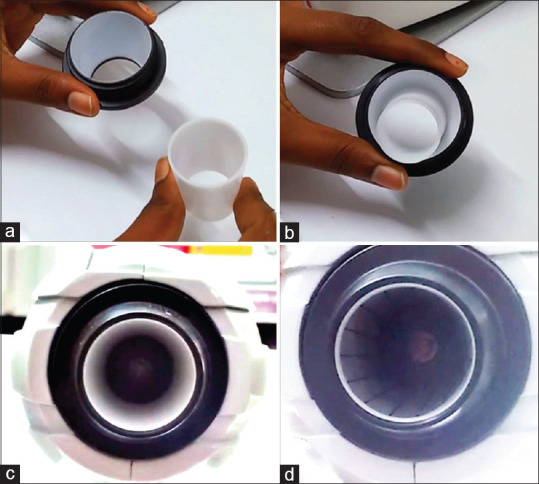
Image showing the (a) various parts of the cone (black and white, respectively), (b) the assembled cone, (c) cone placed in the Tearscope instrument, and (d) Tearscope film inserted into the white cone for a dry eye evaluation
Though this innovation has proved its need and usefulness, there are challenges faced in the Tearscope films, which we would like to shed light upon. Considering that the image of the grid is the end point of analysis, any distortion or irregularity in the Tearscope film may yield false-positive results hampering the tear film assessment. Eroded mires in the NIBUT grid films may go unnoticed if the designated protocols for changing and assessing the films are overlooked.
To avoid such mishaps, it is imperative to have regular and timely assessments to look for eroded grid films. Protocol for timely change of the NIBUT grid films should be implemented. The assessor should prudently look for distortion of mires secondary to an eroded grid, rather than tear film abnormalities [Fig. 2 and Video 2], in which case, the grid should be replaced and the test should be repeated. Currently, the most possible explanation for grid erosion is the friction between the strip and the fingers of the investigator while loading it. Depending on the handling, the strips can last from 1 to 5 months.
Figure 2.
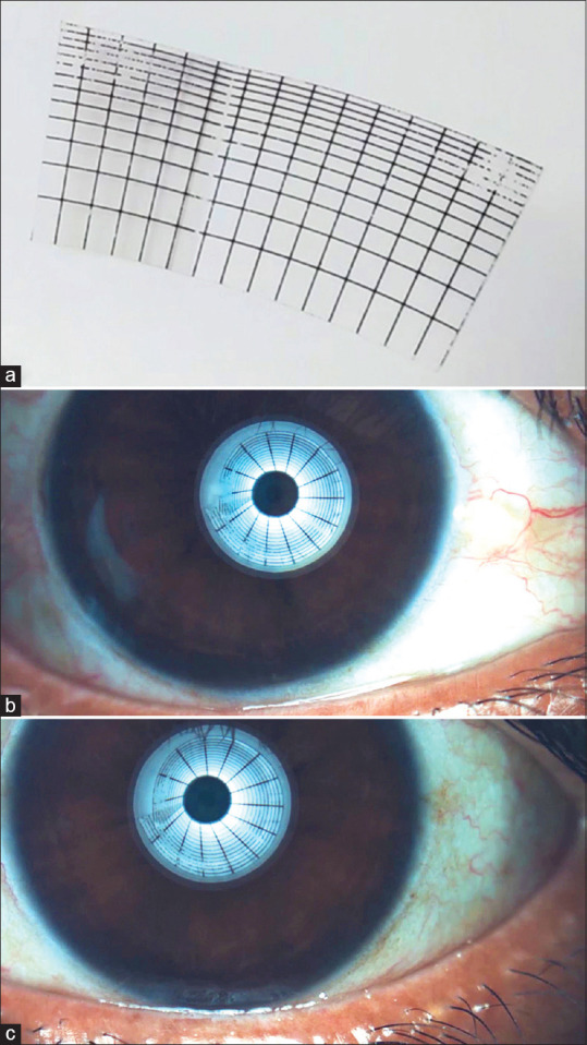
Image showing the (a) Tearscope film used for NIBUT and (b and c) distorted mires of the right eye and left eye, respectively, due to an eroded Tearscope film. NIBUT = noninvasive breakup time
The Tearscope provides the specialist with noninvasive information on the structure and conduct of the tear film, and hence, it can be cumulatively used for preoperative evaluation in ocular surface surgeries.[1,5] Manufacturers recommend the advanced updated version of the Tearscope, where the grid films are inbuilt inside the cone, rather than manually setting it up every time, causing friction to the grid film resulting in segmental loss of mires and false-positive results. Or conversely, users with the traditional model will have to implant frequent grid film inspection regulations to make it a worthy addition to their noninvasive dry eye tool kit.
Financial support and sponsorship
Nil.
Conflicts of interest
There are no conflicts of interest.
Videos avaliable on: www.ijo.in
References
- 1.Koh S, Rao S, Srinivas S, Tong L, Young A. Evaluation of ocular surface and tear function-A review of current approaches for dry eye. Indian J Ophthalmol. 2022;70:1883–91. doi: 10.4103/ijo.IJO_1804_21. [DOI] [PMC free article] [PubMed] [Google Scholar]
- 2.Markoulli M, Duong TB, Lin M, Papas E. Imaging the tear film: A comparison between the subjective Keeler Tearscope-PlusTM and the objective Oculus®Keratograph 5M and LipiView®interferometer. Curr Eye Res. 2018;43:155–62. doi: 10.1080/02713683.2017.1393092. [DOI] [PubMed] [Google Scholar]
- 3.Masmali AM, Purslow C, Murphy PJ. The tear ferning test: A simple clinical technique to evaluate the ocular tear film. Clin Exp Optom. 2014;97:399–406. doi: 10.1111/cxo.12160. [DOI] [PubMed] [Google Scholar]
- 4.Tong L, Lim ST. Review of literature on measurements of non-invasive break up times, lipid morphology and tear meniscal height using commercially available hand-held instruments. Curr Eye Res. 2018;43:567–75. doi: 10.1080/02713683.2018.1437454. [DOI] [PubMed] [Google Scholar]
- 5.García-Marqués JV, Martínez-Albert N, Talens-Estarelles C, García-Lázaro S, Cerviño A. Repeatability of non-invasive keratograph break-up time measurements obtained using oculus keratograph 5M. Int Ophthalmol. 2021;41:2473–83. doi: 10.1007/s10792-021-01802-4. [DOI] [PubMed] [Google Scholar]
Associated Data
This section collects any data citations, data availability statements, or supplementary materials included in this article.