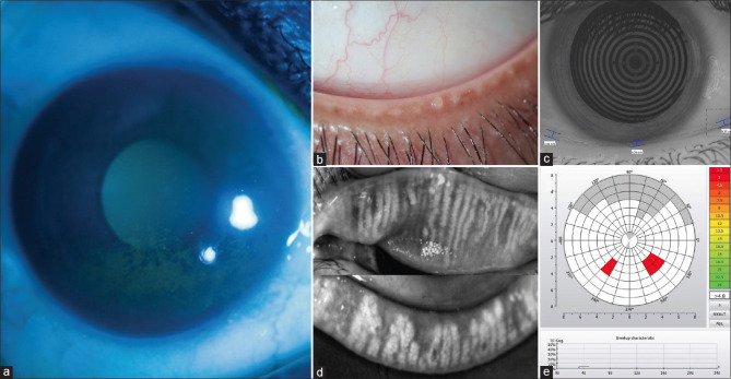
Figure 1.
(a) Typical pattern of corneal fluorescein staining seen under cobalt blue illumination on slit-lamp biomicroscopy in the inferior quadrant adjacent to the lid margin in a case of evaporative dry eye due to meibomian gland dysfunction. (b) Pouting and blocked meibomian gland orifices along the lower lid margin. (c) Reduced tear meniscus height (TMH) as measured by the Keratograph. (d) Infrared imaging of the lids shows areas of meibomian gland dropouts, gland distortion or tortuosity, and gland shortening. (e) Non-invasive-Keratograph-break-up time (NIKBUT) showing abnormal value and pattern

