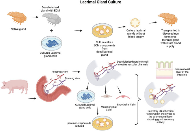
Figure 5.
Above is a schematic representation of tissue-based lacrimal gland reconstruction- Extracellular matrix obtained from the decellularised lacrimal gland is repopulated with primary lacrimal gland cells to generate lacrimal gland constructs which are transplanted into the diseased gland with intact blood supply. Below is a schematic representation of developing a vascularised Lacrimal gland construct/spheroid/organoid grown within a segment of the porcine small intestine (jejunum) with intact vascular channels stemming from the iliac artery and veins. The cultured primary lacrimal gland cells along with MSCs and endothelial cells are grown in the submucosal region of the crypts of the intestine. These vascular channels can be connected to a bioreactor with perfusion of culture media mediated through pressure sensors and peristaltic pumps to maintain the nutrition of the constructs and develop viable blood vessels

