
Fig. 2.
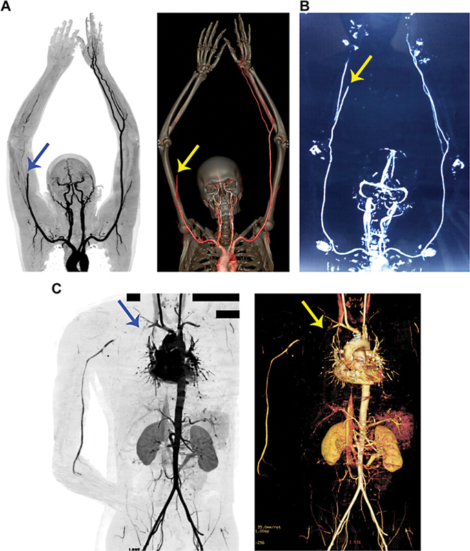
CT angiography reveals occluded peripheral arteries and a lack of downstream blood flow in Russell's bite victims. ( A ) The 2D contrasting and 3D constructed images of CT angiography confirm the occlusion of the right brachial artery and the lack of blood flow to downstream arteries in the first patient. ( B ) The 2D inverted contrasting image of CT angiography confirms the occlusion of the radial artery and blockade of blood flow downstream in the hand and fingers in the second patient. Similarly, 2D and 3D CT angiography ( C ) images confirm the occlusion of the right subclavian artery and the affected downstream blood flow in the third patient. The arrows indicate the site of occlusion. The black rectangle boxes are used to hide personal details on the image.