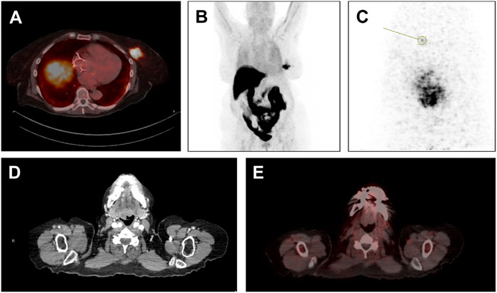
Fig. 2.
18F-FES imaging in patient with left breast ER-positive HER2-negative ILC and oropharyngeal mass for which nondiagnostic biopsy had been performed. (A) Shows left breast mass with 18F-FES uptake on fused PET-CT reflecting ER positivity. (B) Shows 18-F PET highlighting tumor in left breast, with expected uptake of 18F-FES in liver and gastrointestinal tract. In (C), left breast is imaged with dedicated breast PET using 18-F FES, identifying a possible satellite lesion anterior to known tumor. Finally, (D) shows image from CT scan demonstrating irregular oropharyngeal mass, and fused image from 18-F FES PET-CT (E) shows no uptake in mass, suggesting that this mass was unrelated to primary ILC tumor.

