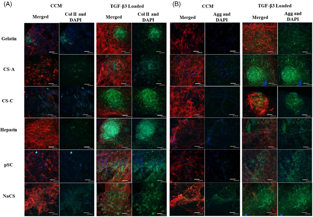
FIGURE 4.
Confocal images of cells cultured on gelatin and gelatin scaffolds containing native GAGs and cellulose sulfate without TGF-β3 (CCM−) or with TGF-β3 loading (TGF-β3 loaded) for 28 days in CCM−. Immunostaining for (A) collagen type II (green) and (B) aggrecan (green). Actin (red) and nucleus (blue) for both A and B panels (40× magnification, scale bar is 50 μm)

