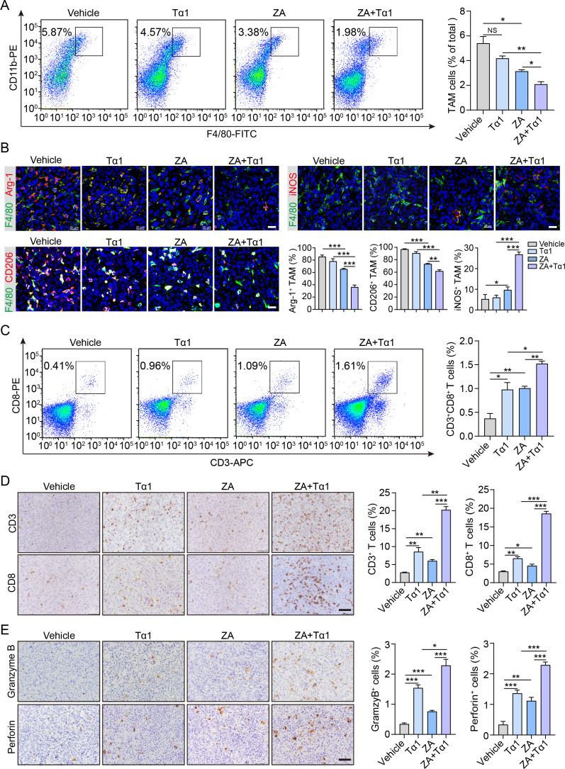
Figure 3.
ZA plus Tα1 therapy enhances tumor inflammation and cytotoxic T-cell infiltration in PCa tumors. (A) Flow cytometric analysis was performed to determine the frequency of CD11b+/F4/80+ macrophages in PCa tumors treated as indicated. (B) IF staining and the proportional changes in M2 macrophages (F4/80+CD206+ or F4/80+Arg-1+) and M1 macrophages (F4/80+ iNOS+) in PCa tumors receiving treatment with vehicle, ZA, Tα1, or ZA+Tα1. Scale bar: 20 µm. Quantification of the IF staining is shown. (C) Flow cytometric analysis was used to evaluate the infiltration of CD3+ and CD8+ T cells in PCa tumors treated as indicated. (D) IHC staining and quantification of infiltrated CD3+ and CD8+ T cells in PCa tumors treated with vehicle, ZA, Tα1, or ZA+Tα1. Scale bar: 50 µm. Quantification of the IHC staining is shown. (E) IHC staining and quantification of the levels of cytotoxic effector molecules (granzyme B and perforin) in PCa tumors treated as indicated. Scale bar: 50 µm. Data are presented as the mean±SEM. n=5. *P<0.05, **P<0.01, and ***P<0.001. NS, no significance. IF, immunofluorescence; IHC, immunohistochemical; PCa, prostate cancer; TAM, tumor-associated macrophage; Tα1, thymosin α1; ZA, zoledronic acid.

