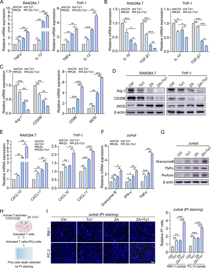
Figure 5.
ZA and Tα1 treatment induces pro-inflammatory macrophages and promotes cytotoxic T cells in vitro. (A, B) RAW264.7 and THP-1 cells were treated with ZA and/or Tα1 for 12 hours, and qPCR was performed to determine the expression levels of (A) TNF-α, IL-12, (B) IL-10 and TGF-β1. (C, D) The expression of markers associated with M2 (Arg-1 and CD206) and M1 (CD86 and iNOS) polarization in macrophages treated with ZA and/or Tα1 for 24 hours was determined by (C)qPCR and (D) Western blotting. (E) qPCR analysis of the expression of CXCL10 and CXCL11 in RAW264.7 and THP-1 cells after the indicated treatments. (F, G) The expression of T-cell cytotoxic effector molecules (granzyme B, TNF-α, IFN-γ, and perforin) in Jurkat cells treated with ZA and/or Tα1 for 24 hours was determined by (F) qPCR and (G) Western blotting. (H) Jurkat cells were activated with anti-CD3/CD28 and then treated with ZA and/or Tα1. Then, the Jurkat cells were co-cultured with RM-1 and PC-3 cells for 24 hours. (I) A PI staining assay was conducted to evaluate the cytotoxic effect of treated Jurkat cells on PCa cells. Representative images and quantification of PI-stained cells are shown. Data are presented as mean±SEM. n=3. *P<0.05, **P<0.01, and ***P<0.001. IFN, interferon; IL, interleukin; mRNA, messenger RNA; PCa, prostate cancer; PI, propidium iodide; TGF, transforming growth factor; TNF, tumor necrosis factor; Tα1, thymosin α1; ZA, zoledronic acid.

