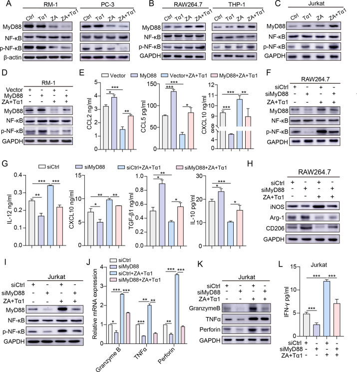
Figure 6.
The MyD88/NF-κB signaling is required for ZA plus Tα1 therapy-mediated immunostimulatory functions of PCa cells, macrophages, and T cells. (A) RM-1 and PC-3 cells, (B) RAW264.7 and THP-1 cells, and (C) Jurkat cells were treated with ZA (25 µM) and/or Tα1 (60 µg/mL) for 6 hours. The levels of MyD88, NF-κB, and p-NF-κB (Ser536) were determined by Western blotting. GAPDH served as a loading control. (D, E) RM-1 cells were transfected with a vector or MyD88 overexpression plasmid for 48 hours, followed by treatment with ZA and Tα1 for 6 hours. Then, (D) the expression of MyD88, NF-κB and p-NF-κB (Ser536) was evaluated by Western blotting, and (E) the expression of CCL2, CCL5, and CXCL10 in RM-1 cells was determined by ELISA. (F) RAW264.7 cells were transfected with NC siRNA (siCtrl) or MyD88-specific siRNA (siMyD88) for 24 hours, followed by treatment with ZA and Tα1 for 6 hours. Then, the expression of MyD88, NF-κB and p-NF-κB (Ser536) was determined by western blotting. (G) ELISA analysis of the levels of IL-12, CXCL10, TGF-β1, and IL-10 produced by RAW264.7 cells in each group. (H) Western blot analysis of the expression of iNOS, Arg-1, and CD206 in macrophages treated and transfected as indicated. (I) Jurkat cells were transfected with NC siRNA or MyD88-specific siRNA for 24 hours, followed by treatment with ZA and Tα1 for 6 hours. The expression of MyD88, NF-κB and p-NF-κB (Ser536) was determined by Western blotting. (J, K) The expression of granzyme B, TNF-α, and perforin was determined by (J) qPCR and (K) Western blotting. (L) The expression of IFN-γ by T cells from each group was evaluated by ELISA. Data are presented as mean±SEM. n=3. *P<0.05, **P<0.01, and ***P<0.001. IFN, interferon; IL, interleukin; mRNA, messenger RNA; PCa, prostate cancer; siRNA, small interfering RNA; TGF, transforming growth factor; TNF, tumor necrosis factor; siCtrl, negative control siRNA; Tα1, thymosin α1; ZA, zoledronic acid.

