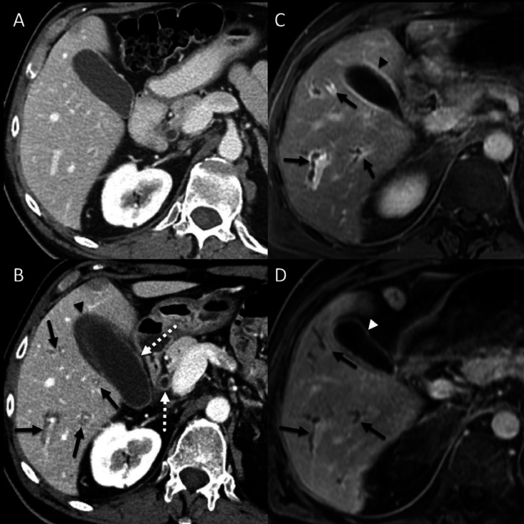
Figure 2.
A patient with non-small cell lung cancer. (A) Baseline contrast-enhanced CT image showing unremarkable appearance of the liver. (B) Contrast-enhanced CT image at about 1 year after immune checkpoint inhibitor (ipilimumab and nivolumab) initiation showing periportal cuffing (solid arrows), thickening and avid enhancement of the common bile duct and the gallbladder wall (dotted arrows) and pericholecystic fluid (arrowhead). (C) 5-min delayed post-contrast gradient spoiled echo image showing periportal cuffing (arrows) and avid enhancement of the gallbladder wall (arrowhead), these findings are consistent with cholangitis. (D) 5-min delayed post-contrast gradient spoiled echo image 3 months after initiation of anti-IL-6R therapy showing resolution of periportal enhancement but there is persistent irregularity and mild dilatation of the intrahepatic biliary ducts (arrows). Avid enhancement of the gallbladder wall has also resolved (arrowhead). anti-IL-6R, anti-interleukin-6 receptor; ICI, immune checkpoint inhibitor.

