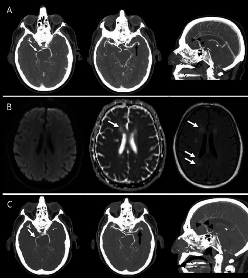
Figure 3.
A patient with malignant melanoma. (A) Maximal intensity projection (MIP) images of concurrent cranial CT angiography at 7 months after immune checkpoint inhibitor (ipilimumab and nivolumab) initiation show multiple sites of intracranial arterial narrowing and/or occlusion involving the right posterior cerebral artery (white arrow), left middle cerebral artery and both anterior cerebral arteries (black arrowheads). This multi vascular pattern of involvement is suggestive of vasculitis. (B) Diffusion-weighted image, corresponding apparent diffusion coefficient (ADC) map, and fluid-attenuated inversion recovery (FLAIR) 3 months after initiation of anti-IL-6R therapy show normalization of ADC signal and development of high signal intensity foci on FLAIR (arrows) without corresponding diffusion restriction, in keeping with chronic infarcts. (C) MIP images of cranial CT angiography after anti-IL-6R therapy show recanalization with persistent narrowing of the right posterior cerebral artery (white arrow), persistent narrowing of the left middle cerebral artery, and recanalization of the anterior cerebral arteries (black arrowheads). anti-IL-6R, anti-interleukin-6 receptor.

