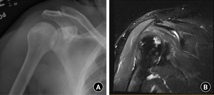
Fig. 2.
Preoperative plain anterior-posterior with internal rotation and T2-weighted coronal magnetic resonance imaging (MRI) of the shoulder showing stage I avascular necrosis of the humeral head. (A) The plain radiographic view of the shoulder showing absent osteosclerosis or articular abnormality. (B) T2-weighted MRI showing a T2 signal intensity of the medial humeral head, consistent with osteonecrosis.

