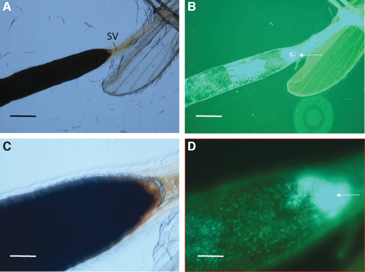
FIG. 4.
Micrographs of an isolated sand fly anterior midgut at 5 days postfeeding. The anterior portion of the anterior midgut is above the wing. (A) The blood meal was a dark, rusty color by phase-contrast microscopy (100 × magnification; scale bars = 160 μm). The SV is indicated. (B) Corresponding fluorescence micrograph revealing GFP+ B. ancashensis cells in the lumen of the anterior midgut with a microcolony at the anterior end of the anterior midgut just below the SV (arrowed). (C) Closeup image of (A) phase-contrast microscopy (400 × magnification; scale bars = 50 μm). (D) Corresponding fluorescence micrograph to (C), revealing GFP+ B. ancashensis cells just below the SV.

