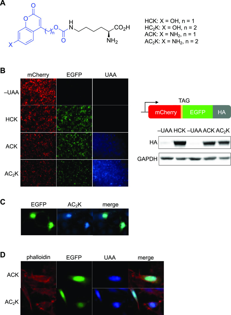
Figure 1.
Incorporation of ACK and AC2K into proteins in mammalian cells. (A) Chemical structures of HCK, HC2K, ACK, and AC2K. The photolabile group/fluorophore is colored blue. (B) Confirmation of ACK and AC2K incorporation into a reporter construct through fluorescence imaging in HEK293T cells (10× magnification) and western blot. (C) Expression of NLS-EGFP-AC2K in live NIH 3T3 cells (20× magnification). (D) Fixed HeLa cells expressing NLS-EGFP-ACK (top row, 40×) and NLS-EGFP-AC2K (bottom row, 63×). Cells are counterstained with rhodamine–phalloidin.

