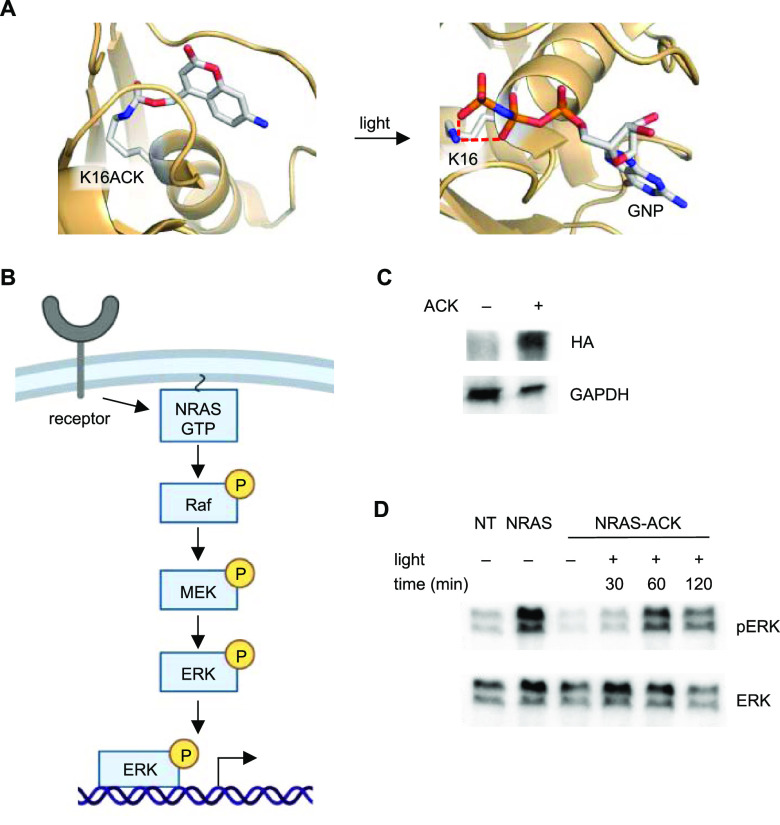
Figure 4.
Optical control of a RASopathy mutant of NRAS. (A) Structural representation of the caged NRAS nucleotide-binding site before and after light exposure (models generated from PDB: 5UHV). (B) Diagram of the general RAS/MAPK signaling pathway. (C) Western blot of ACK incorporation into HA-NRAS G60E K16TAG (NRAS-ACK). (D) Western blot of phospho-ERK at different timepoints after irradiation of NRAS-ACK. NT = nontreated embryos.

