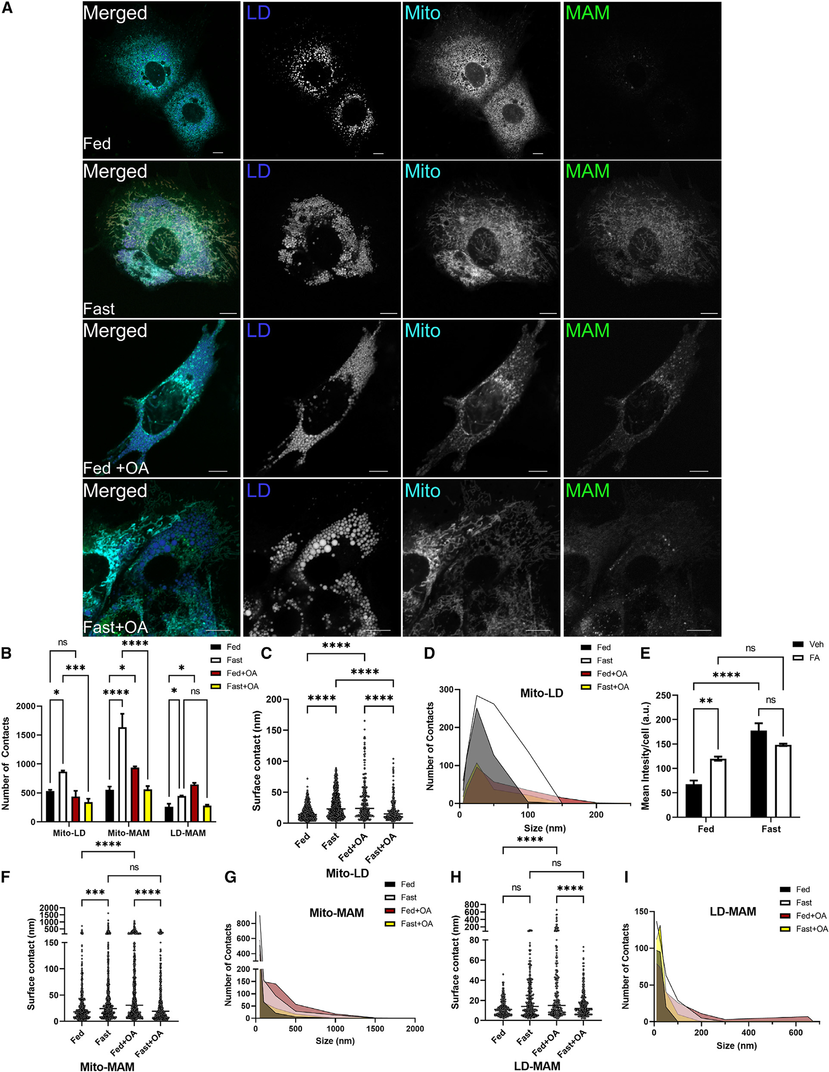
Figure 6. FAs regulate mitochondrion-LD and mitochondrion-ER-LD contacts regardless of metabolic state.

(A) Representative confocal microscopy images of mitochondria and LDs in AML12 cells in fed, fasting, fed plus 250 mM oleate, or fasting plus 250 mM oleate in the medium.
(B) DiAna plug-in colocalization determined from images in (A), quantifying the number of MITO-LD, MAM-MITO, and MAM-LD contacts as determined by the DiAna plug-in.
(C) Surface contact length between mitochondria and LDs in the fed and fasted state.
(D) Histogram outlining the change in contact length determined in (C).
(E) Quantification of SPLICS by total mean intensity per cell. n = 6–8 images with 12–16 cells per condition.
(F) Surface contact length between SPLICS and mitochondria in the fed and fasted state as determined by the DiAna plug-in in FUJI.
(G) Histogram outlining the change in contact length determined in (F).
(H) Quantification of surface contact length between SPLICS and LDs in fed and fasted states.
(I) Histogram outlining the change in contact length determined in (H).
ns, p > 0.05; *p < 0.05; **p < 0.01; ***p < 0.001; ****p < 0.0001.
