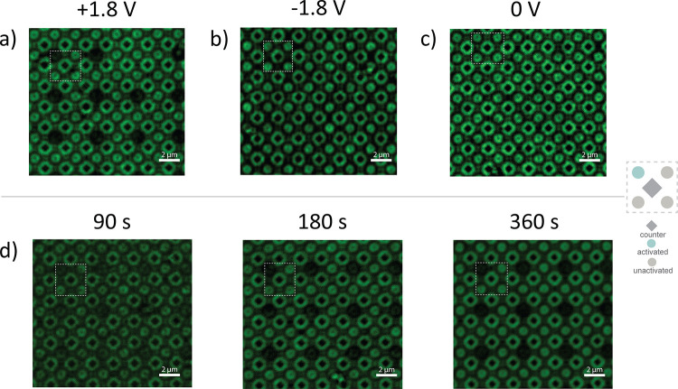
Figure 5.
Cleavage of conjugate 5 addition product. dT initiators were extended with conjugate 5 and a cell potential applied to one of the four groups of working electrodes. The array surfaces were then stained with 6x-His antibody. Uniform fluorescent intensity is indicative of the presence of conjugate 5 across the surface, and a lack of fluorescence is indicative of the removal of conjugate 5 from that region. (a) +1.8 V was applied to single working electrode for 90 s. (b) −1.8 V was applied to single working electrode for 90 s. (c) The array was incubated in cleavage buffer for 90 s with no potential applied. (d) +1.0 V was applied to a single working electrode for the indicated time.

