
Figure 4.
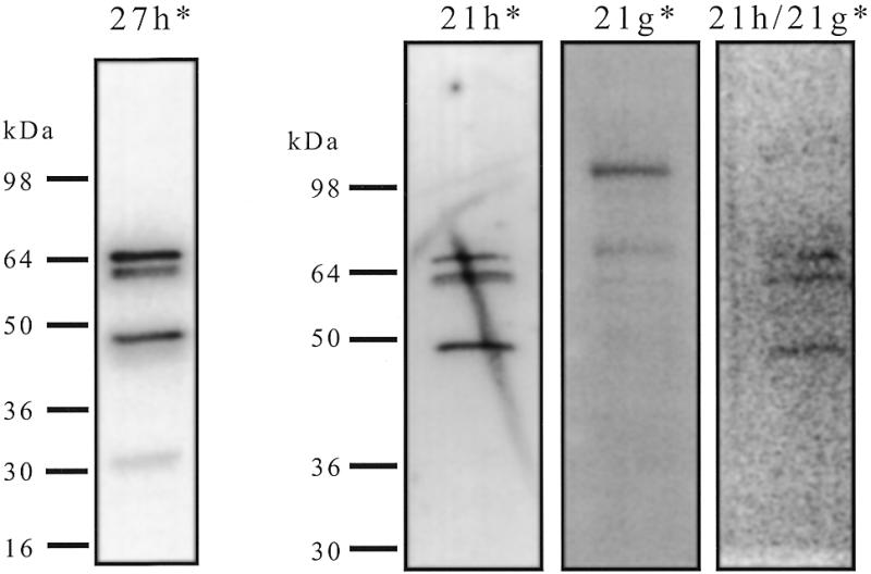
Southwestern analysis of HNE with different probes. Aliquots of 4.5 µg of HNE were run on an 8–16% SDS–PAGE gel and transferred onto a PVDF membrane. After blocking and renaturation each strip of the membrane was hybridised with a different radiolabelled probe (0.4 nM) (27h*, 21h*, 21g* or a preformed duplex 21h/21g*) in HEPES buffer pH 7.2, in the presence of single- and double-stranded competitors, washed three times and exposed on a phosphorimager screen. *, the radiolabelled strand. The position of molecular weight markers was obtained using coloured markers transferred to the membrane.