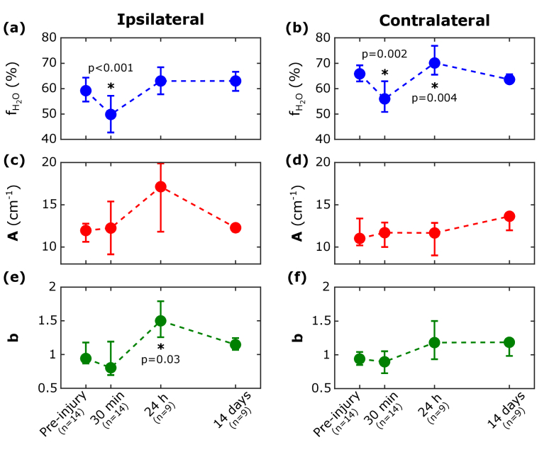
Fig. 4.
Pre-injury and post-injury diffuse optical measurements of cerebral tissue water volume fraction , scattering amplitude (A), and scattering power (b) for the ipsilateral and contralateral hemispheres (A and b are defined by Eq. (1)). The medians (circles) and interquartile ranges across subjects for each measurement timepoint are shown. P values indicate whether the median pre-injury to post-injury change was different from zero.

