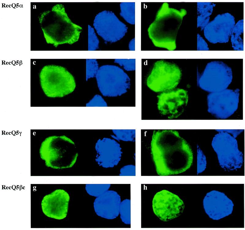
Figure 5.
Subcellular localization of RecQ5 isoforms in 293EBNA cells. Each construct was transiently expressed in 293EBNA cells and the cells fixed and stained with anti-HA antibody as shown in Materials and Methods. Localization of the RecQ5 isoform and RecQ5βc proteins are indicated by FITC (green, left panel) and the nuclear positions are shown by DAPI (blue, right panel). (a and b) RecQ5α; (c and d) RecQ5β; (e and f) RecQ5γ; (g and h) RecQ5βc.

