
Figure 2.
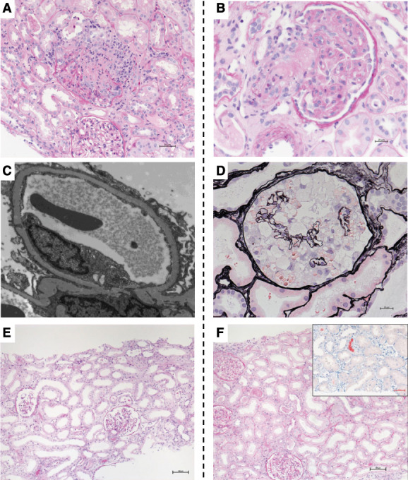
Representative kidney biopsy findings of postvaccination case and those with COVID-19. The figure illustrates representative kidney histopathology findings in patients after SARS-CoV-2 vaccination (panels A, C, E) or with COVID-19 (panels B, D, F). (A) Light microscopy image showing ANCA-associated necrotizing GN and ATI in case V1. PAS staining. Magnification, ×200. (B) Light microscopy image showing TMA in case C7. PAS staining. Magnification, ×200. (C) Diffuse foot process effacement (minimal changes) in case V11. Transmission electron microscopy. Magnification, ×8000. (D) Light microscopy image showing a collapsing glomerulopathy lesion characterized by glomerular epithelial cell hyperplasia and underlying glomerular capillary collapse in case C8 of African ancestry. Jones methenamine silver staining. Magnification, ×400. (E) Light microscopy image showing ATI with dilatation, flattened epithelium, and brush border defects in case V21 that had ATI. PAS staining. Magnification, ×100. (F) Light microscopy image showing ATI and myoglobin casts (inset shows myoglobin immunohistochemistry staining) in case C12. PAS. Magnification, ×100. PAS, periodic acid–Schiff.