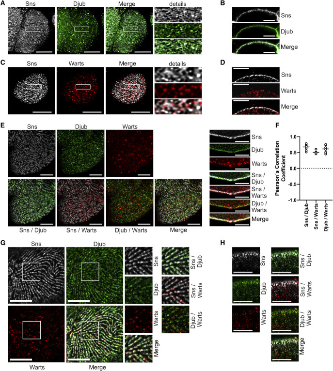
Figure 1.
Djub and Warts colocalize with Sns at nephrocytes SD. (A) Microscopy of wild-type third instar larvae garland nephrocytes stained with antibodies against Sns (gray) and Djub (green) after PFA fixation. The staining with Sns shows the typical fingerprint-like structure of SDs on the nephrocyte cortex. Djub staining shows a very similar structure with partial colocalization of Sns and Djub at SD. (B) Cross-sections of nephrocytes as used in (A) confirm partial colocalization of Sns and Djub at the cortex. (C) Microscopy of third instar larvae garland nephrocytes overexpressing myc-Warts stained with antibodies against Sns (gray) and myc (red) after fixation in a heat fix solution. Warts staining partly overlapped with the typical fingerprint-like staining pattern, confirming partial SD localization of Warts. (D) Cross-sections of nephrocytes as used in (C) proved partial SD localization of Warts. An additional staining of Warts was detected within the cytoplasm of the nephrocytes, which may be an effect of myc-WartsOE. (E) Microscopy of third instar larvae garland nephrocytes overexpressing GFP-Djub and myc-Warts stained with antibodies against Sns (gray), GFP (green), and myc (red) show colocalization between Djub and Sns in the fingerprint-like staining pattern on the nephrocyte surface (left). Warts staining at SD is less than Djub staining but, where detected, Warts colocalizes with Djub and Sns. Cross-sections through the nephrocytes confirm that Djub strongly and Warts partly colocalizes with Sns at SD (right). When overexpressed, Djub and Warts additionally localize within the cytoplasm. In the case of Djub, this is an effect of overexpression (compare B). Lack of Warts-specific antibody makes this proof impossible for Warts. (F) Evaluation of colocalization between the proteins Sns and Djub, Sns and Warts, and Djub and Warts by determination of Pearson coefficients confirmed strong correlation between localization of all three proteins (mean values between 0.5 and 1). (G) Expansion microscopy of nephrocytes as used in (E). Surface analyses confirm very similar structures and partial colocalization between Djub and Sns at SD. Warts also colocalizes with SD structures. (H) Cross-sections of nephrocytes as shown in (E) using expansion microscopy confirm colocalization of Sns and Djub at SD and, to a smaller amount, colocalization with Warts. Scale bars (A–F) 5 µm and (G and H) 20 µm. Figure 1 can be viewed in color online at www.jasn.org.

