
Figure 5.
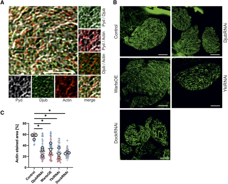
Djub connects the SD of nephrocytes with the cytoskeleton. (A) Immunostaining of third instar larvae garland nephrocytes with antibodies against Djub (green) and Pyd (gray). F-actin was stained using phalloidin (red). Details show actin filaments crossing Djub and Pyd-stained SDs or partial colocalization at intersections. (B) Actin staining with phalloidin (green) of third instar larvae garland nephrocytes after knockdown of Djub, Yki, or Dock (DjubRNAi, YkiRNAi, DockRNAi) or Warts overexpression showed a reduction of actin strains on the surface. (C) Densitometric quantification of F-actin by measurement of the relative coverage on the surface, normalized to control, illustrates this reduction caused by DjubRNAi, YkiRNAi, DockRNAi, or Warts overexpression (n>30). Scale bars 5 µm. Figure 5 can be viewed in color online at www.jasn.org.