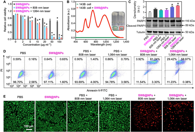
Fig. 4.

Photothermal effect of SW8@NPs under laser irradiation in vitro. (A) Viabilities of 143B cells at diverse concentrations of SW8@NPs irradiated by 808-/1,064-nm lasers (0.23 W cm−2) (n = 6, *P < 0.05 vs. SW8@NPs). (B) Cellular uptake of SW8@NPs to 143B cells. (C) Representative images (down) and quantification (up) of Western blots against PARP1, cleaved-PARP1, and tubulin after various treatment (n = 3, *P < 0.05 vs. PBS). (D) Cell apoptosis of 143B cells examined by flow cytometry. (E) Fluorescence imaging of 143B cells (96-well plates) stained with calcein AM and ethidium homodimer-1 (green: live cells, red: dead cells). Scale bar = 100 μm.
