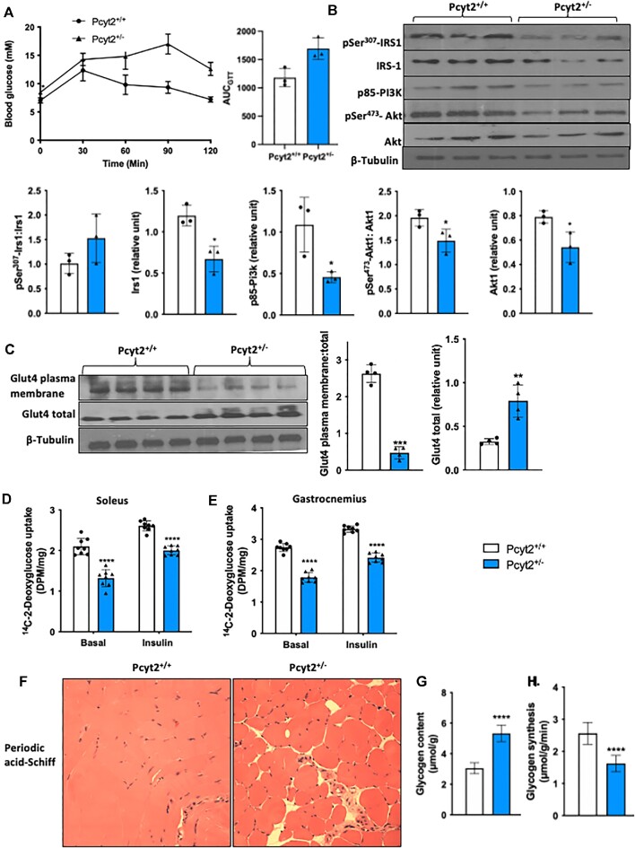
Figure 5.
Impaired glucose transport and glycogen accumulation. (A) Glucose tolerance test and area under the curve showing whole-body reduced insulin sensitivity in Pcyt2+/- relative to Pcyt2+/+. (B) Western blot analysis of proteins in the insulin signaling cascade: pSer307-Irs1, Irs1, p85-Pi3k, pSer473-Akt, Akt; pSer307-Irs1 relative to Irs1; pSer473-Akt relative to Akt; and Irs1, p85-Pi3k, and Akt normalized to β-Tubulin show reduced expression, indicating impaired insulin signaling in Pcyt2+/- whole muscle. (C) Western blot analysis of plasma membrane Glut4 content and total Glut4 content; Glut4 plasma membrane relative to Glut4 total, and Glut4 total normalized to β-Tubulin. This shows reduced activation and translocation of Glut4 to the plasma membrane in Pcyt2+/- whole muscle. 14C-2-deoxyglucose uptake into (D) soleus and (E) gastrocnemius muscles with and without insulin stimulation also showed diminished glucose uptake in Pcyt2+/−. (F) Periodic acid-Schiff staining of quadricep muscle sections showing glycogen accumulation, and (G) assay-based quantification showing elevated levels of glycogen content in Pcyt2+/- whole muscle. (H) Rate of glycogen synthesis is decreased in Pcyt2+/- whole muscle. Together, this shows impaired glucose handling in Pcyt2+/- with glycogen accumulation that inhibits further synthesis, insulin signaling and glucose update. Data are derived from both male and female mice and are presented as mean ± SD. *P < 0.05; **P < 0.01; ***P < 0.001; ****P < 0.0001.

